
What “dilated” means
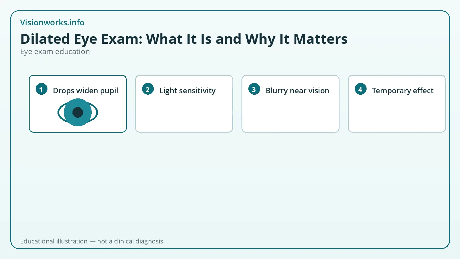
During a dilated eye exam, the clinician places medicated drops that temporarily widen (dilate) your pupils. A wider pupil lets more light enter and gives a broader view of the retina, macula, optic nerve, and blood vessels at the back of the eye.
NEI consumer education describes dilated exams as a key way to look for eye diseases early — when treatment is often more effective — including diabetic retinopathy, glaucoma-related changes, and age-related macular degeneration themes.
Dilation is a tool within a comprehensive exam, not a separate mysterious procedure. You may still have acuity testing, refraction, pressure checks, and anterior segment evaluation in the same visit.
What clinicians look for
With the pupil enlarged, the clinician can better inspect the optic nerve for cupping or pallor patterns that raise glaucoma concern, the macula for aging or degenerative changes, and retinal vessels for diabetes- or hypertension-related signs. Peripheral retina evaluation can be important when patients report flashes, floaters, or have risk factors for tears or detachment.
MedlinePlus materials on standard eye exams note that drops allow ophthalmoscopy of the fundus — retina and nearby structures. AAO exam basics similarly describe dilation as the step that helps examine the retina and optic nerve for disease-related damage.
Findings are interpreted in context: age, symptoms, medical history, prior images, and other test results. A single observation is a data point, not a DIY label you should assign from a web article.
What it feels like
Drops may sting briefly. Your near vision often becomes blurry and outdoor light may feel harsh. These effects are expected and temporary for most people. Bring sunglasses and consider a ride home, especially if you are unsure how you respond.
While dilated, reading phones and paperwork can be frustrating. That inconvenience is the tradeoff for a better internal view. If your job requires immediate precision near work, schedule accordingly.
Rarely, dilating drops can contribute to an acute angle-closure crisis in susceptible eyes. Sudden severe pain, nausea, and marked vision reduction after drops need urgent care. Mention known narrow angles or prior warnings from clinicians.
Dilation, imaging, and “skipping drops”
Some clinics offer widefield retinal photography or other imaging. Images can document findings and educate patients. They may reduce the need for dilation in selected low-risk scenarios — but that is a clinical decision, not a universal rule you should demand based on convenience alone.
If you decline dilation, ask what portions of the retina were not fully evaluated and when the next opportunity for a dilated view should occur. Informed refusal is different from assuming an undilated glance equals a complete posterior exam.
People with diabetes, higher myopia, prior retinal problems, new flashes/floaters, or unexplained vision loss are examples of situations where thorough posterior evaluation is especially important. Your clinician individualizes the plan.
Aftercare and follow-up
Use sunglasses outdoors until pupils recover. Avoid assuming you can safely drive if glare and focus are impaired — arrange alternatives when needed. Resume contacts only when advised if drops or ointment were used.
If the exam finds a new issue, you may receive monitoring intervals, treatment, or referral. If everything looks stable, you still benefit from knowing your baseline. Ask for a plain-language summary of retina and optic nerve status.
Educational symptom pages on Visionworks.info can help you describe floaters or blur at future visits, but sudden curtains of vision loss or dense new floater showers with flashes deserve urgent clinical attention rather than online self-triage.
Deeper context for careful readers
This page (/eye-exams/dilated-eye-exam) is written for education, not diagnosis. The goal is to help you ask better questions at a real appointment and to understand vocabulary you may see on charts, portals, or prescription printouts.
Authoritative organizations such as the AOA, AAO, NEI, and MedlinePlus publish consumer-facing explanations that clinicians still individualize. If anything you read here conflicts with advice from your eye doctor, follow your clinician — they can see your eyes and history; a website cannot.
Between visits, note changes by eye, by distance (far versus near), and by timing (constant versus end of day). Those details make exams more efficient without turning self-tracking into self-diagnosis.
Urgent warning signs still override any routine schedule: sudden vision loss, a curtain or shadow, painful red eye with nausea, chemical exposure, or trauma require prompt in-person care.
Visionworks.info tools can reinforce concepts — acuity notation, prescription fields, symptom vocabulary — while remaining explicitly non-diagnostic. Use them as study aids alongside professional care, not as replacements for refraction, dilation, or medical treatment decisions.
When you compare information across websites, prefer primary sources from AOA, AAO, NEI, MedlinePlus, CDC, FTC, or FDA pages that match your topic. Marketing blogs and unverified social videos often oversimplify intervals, prescriptions, or “pass/fail” chart claims.
Finally, keep expectations proportionate: understanding a term should make your next visit clearer, not turn you into your own optometrist. Curiosity is useful; unsupervised lens ordering from guessed numbers is not.
When professional care matters
Schedule an eye exam if you notice new blur, eye discomfort that persists, headaches tied to visual tasks, difficulty with night driving, or any sudden change in vision. Children, people with diabetes, and anyone with a history of eye disease often need scheduled professional monitoring rather than relying on online tools alone.
Seek urgent or emergency care for sudden vision loss, a curtain or shadow over vision, new flashes with many floaters, severe eye pain, chemical exposure, or eye trauma.