
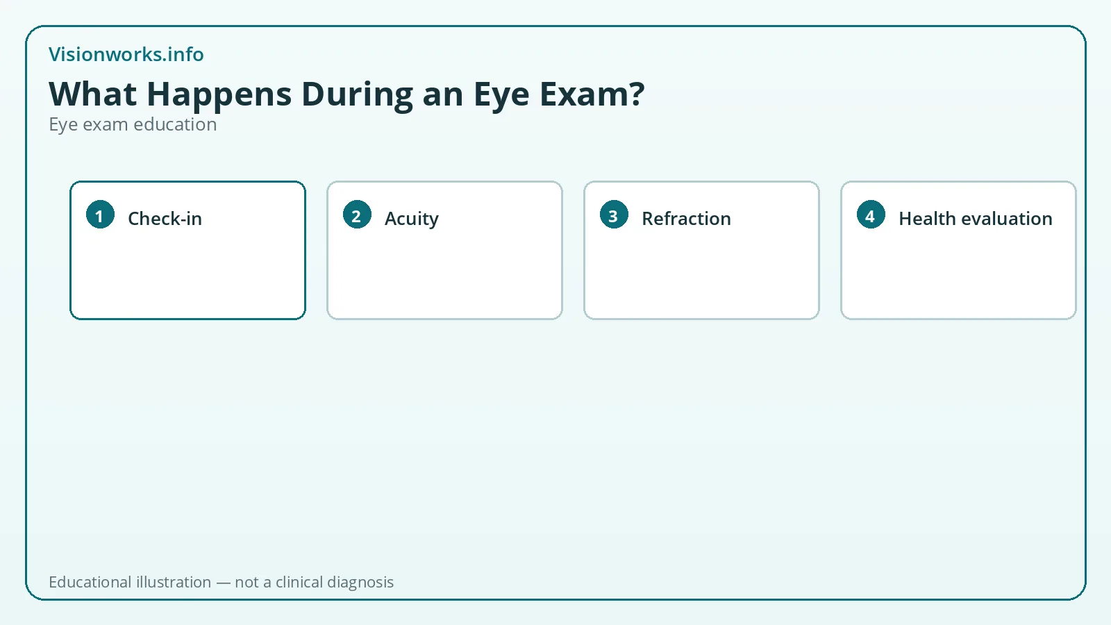
Before you sit in the exam chair
Most visits begin with paperwork or digital intake: the reason for your visit, last exam date, medical and ocular history, allergies, medications, and whether you wear glasses or contacts. Staff may also capture baseline measurements such as blood pressure in some medical eye clinics.
Arrive with your current eyewear. If you wear contacts, ask the office ahead of time whether they want you in lenses or glasses for part of the visit — contact lens evaluations sometimes need both on-eye assessment and over-refraction steps.
Write down symptoms in plain language: which eye, when blur started, distance versus near, glare at night, headaches after screens, or intermittent double vision. Specific descriptions help more than “my vision feels off.”
Visual acuity and preliminary checks
Visual acuity testing is the familiar chart reading. You cover one eye at a time and identify letters, numbers, or symbols at a standardized distance. Near acuity may be tested with a handheld card. Results are recorded as fractions such as 20/20 or 20/40, or in other notations your clinic uses.
Preliminary tests often follow. These can include color vision screening, depth perception, peripheral vision confrontation or automated fields when indicated, pupil responses to light, and eye movement tracking as you follow a target. None of these alone tells the whole story, but together they map function.
AAO patient education describes a comprehensive exam as typically comfortable and often completed within roughly 45 to 90 minutes depending on complexity, dilation, imaging, and whether contact lens fitting is included.
Refraction: “which is better, one or two?”
Refraction determines the lens powers that sharpen focus for refractive errors. An autorefractor may provide a starting estimate. Retinoscopy (a light-and-lens technique) gives an objective clue, especially useful for children or patients who struggle with subjective choices.
Subjective refraction uses a phoropter or trial frame. The clinician alternates lenses and asks which view is clearer. There is no trick question: if both look similar, saying so is useful. Your answers refine sphere, cylinder, and axis for each eye, and an ADD for near when presbyopia is relevant.
Refraction measures focusing need; it is not by itself a complete eye-health exam. Clinicians combine refraction with binocular findings and health results before finalizing a prescription recommendation.
Eye health evaluation
A slit-lamp microscope lets the clinician inspect lids, lashes, cornea, conjunctiva, iris, and the front of the lens under magnification. Fluorescein dye may highlight dry patches or corneal surface issues. Tonometry estimates intraocular pressure as one glaucoma-risk data point — pressure alone does not diagnose glaucoma.
To evaluate the retina, macula, and optic nerve more thoroughly, dilating drops are often used. NEI materials explain that widening the pupil lets more light in so the clinician can examine internal structures for diabetic changes, macular disease, and other problems. Imaging such as retinal photography or OCT may supplement the exam when available and indicated.
If dilated, expect light sensitivity and near blur for several hours. Sunglasses and a ride home are practical planning steps. MedlinePlus standard eye exam education also notes these temporary effects.
Wrapping up: results, prescriptions, and next steps
The clinician should explain key findings in understandable terms: whether refractive error changed, whether eye health looks stable, and what follow-up interval makes sense for you. Ask about anything unclear — especially new medications, home care for dry eye, or why imaging was ordered.
If you need glasses or contacts, you should leave with a clear understanding of what was prescribed and any wearing instructions. Contact lens prescriptions are distinct from glasses prescriptions and typically include brand/material parameters required for safe ordering.
If a finding needs specialty care — for example, a retinal concern — you may receive a referral. That is a coordination step, not a reason to panic by default; many referrals are for confirmation or monitoring.
Staff roles and communication tips
Technicians often gather preliminary data before the doctor reviews results and performs medical decision-making steps. That division of labor is normal and does not mean the exam is incomplete — the licensed clinician remains responsible for interpretation and prescriptions.
If English is not your first language, request an interpreter when scheduling. Misheard lens choices during refraction can produce uncomfortable glasses even when the clinic equipment is excellent.
Ask for a written summary of changes: new sphere values, whether dilation was performed, intraocular pressure readings if taken, and the recommended return date. Paper or patient-portal notes reduce forgotten instructions.
When professional care matters
Schedule an eye exam if you notice new blur, eye discomfort that persists, headaches tied to visual tasks, difficulty with night driving, or any sudden change in vision. Children, people with diabetes, and anyone with a history of eye disease often need scheduled professional monitoring rather than relying on online tools alone.
Seek urgent or emergency care for sudden vision loss, a curtain or shadow over vision, new flashes with many floaters, severe eye pain, chemical exposure, or eye trauma.